
New 3D microscope packs many imaging tools into one system
UTSW-led technology switches among lenses and allows researchers to conduct wider range of experiments, reducing need for multiple pieces of equipment
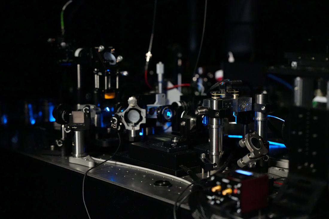
DALLAS – Nov. 05, 2026 – An international team of researchers led by UT Southwestern Medical Center has developed a versatile light-sheet microscope that captures 3D views of biological samples, from activity inside living cells to larger tissues. Designed to make detailed imaging much simpler and faster, the approach could help laboratories stretch equipment budgets and expand research that may ultimately inform patient care.
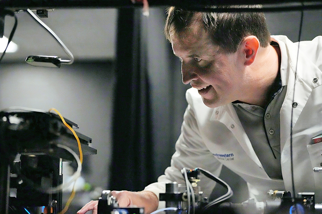
“Our work makes a popular microscope technique, called light-sheet fluorescence microscopy, more versatile and user-friendly,” said corresponding author Reto Fiolka, Ph.D., Associate Professor in the Lyda Hill Department of Bioinformatics and of Cell Biology at UT Southwestern. “It extends what a single instrument can do in terms of sample coverage and imaging applications.”
Described in Nature Methods, the multi-immersion oblique plane microscope (miOPM) addresses a long-standing challenge: Advanced microscopes are often designed for a narrow range of tasks. Studying different samples can require separate instruments, specialized preparation, and considerable technical expertise.
The new platform provides a practical blueprint for making sophisticated 3D imaging available to more researchers, helping more labs pursue questions that existing configurations may place beyond reach. The potential savings come from reducing the need for multiple dedicated systems and the work involved in reconfiguring them, although the study does not quantify financial savings.
Dr. Fiolka led the study with co-first author Bingying Chen, Ph.D., a former Instructor and postdoctoral researcher in the Fiolka Lab who is now a Research Specialist at the Howard Hughes Medical Institute’s Janelia Research Campus. In addition to UTSW and other U.S. institutions, the team included researchers from Australia, Germany, Poland, and the U.K.
Traditional light-sheet microscopes are usually built like single-purpose tools. Some are optimized to zoom in on tiny structures inside a cell, requiring oil on the lens (i.e., an oil immersion objective). Others are designed to look at larger tissues or organoids, requiring water or special fluids. Some need to scan quickly across hundreds of sample dishes without touching them, requiring air between the lens and sample. Switching between these jobs requires labs to buy and maintain multiple expensive microscopes.
The miOPM allows researchers to switch objective lenses on a single microscope designed for use with oil, water, or air, adapting it for various experiments without needing different equipment. An adjustable zoom lens helps preserve image quality when scientists change the liquid surrounding a sample. Together, these features let one system handle a broader range of studies without rebuilding its internal optics.
Like other light-sheet microscopes, miOPM illuminates one thin section of a sample at a time, building a 3D picture while limiting unnecessary light exposure. This approach supports rapid imaging and helps protect living samples from light-related damage, allowing scientists to follow biological activity as it unfolds.
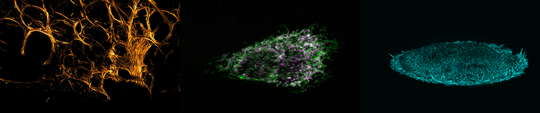
The team demonstrated the platform by observing moving structures on immune cells, tracking activity within cells’ energy-producing structures, and examining lung and liver cancer tissue. The system also worked with standard laboratory plates containing hundreds of sample wells, showing its capability of studying many samples efficiently.
For disease research, this flexibility could help scientists investigate how cells function, how disease changes tissue, and how possible treatments affect biological processes. Over time, these insights could contribute to better diagnostic approaches and therapies.
The new technology’s potential impact on patient health is also significant. Because it can rapidly scan 384-well plates containing live human cells or mini-tumors, pharmaceutical researchers conceivably could test thousands of drug candidates much faster to see how they affect disease targets in 3D. Also, by capturing clear 3D videos of living immune cells, brain cells, and lung organoids, doctors and scientists could uncover the root causes of conditions such as Parkinson’s, cancer, and respiratory diseases, leading to more targeted therapies.
“This project is a great example of how UT Southwestern is creating next-generation scientific technologies that are both high-performing and accessible,” Dr. Fiolka said. “By lowering costs while maintaining advanced capabilities, we can help more researchers accelerate biomedical discovery.”
The imaging in this study was performed with two miOPM systems – one at UT Southwestern and one at Calico Life Sciences in San Francisco.
A complete list of authors from UTSW can be found in the study.
Dr. Fiolka is a member of the Development and Cancer Research Program at the Harold C. Simmons Comprehensive Cancer Center.
This research was supported by the National Institutes of Health, National Institute of General Medical Sciences (R35GM133522, R35GM119768, R35GM137894, and RM1GM145399); the National Institute of Biomedical Imaging and Bioengineering (R01EB035538); the National Institute of Mental Health (DP2MH119423); and the National Cancer Institute (U54CA268072).
About UT Southwestern Medical Center
UT Southwestern, one of the nation’s premier academic medical centers, integrates pioneering biomedical research with exceptional clinical care and education. The institution’s faculty members have received six Nobel Prizes and include 28 members of the National Academy of Sciences, 26 members of the National Academy of Medicine, and 14 Howard Hughes Medical Institute Investigators. The full-time faculty of nearly 3,400 is responsible for groundbreaking medical advances and is committed to translating science-driven research quickly to new clinical treatments. UT Southwestern physicians in more than 80 specialties care for more than 143,000 hospitalized patients, attend to more than 470,000 emergency room cases, and oversee nearly 5.3 million outpatient visits a year.